Author: Reneesh, (Consultant Orthopedic Surgeon – Wellkins Medical Centre)
It is one of the things parents notice early. A toddler taking their first steps with legs that curve outward like a little cowboy. Or a child of five or six whose knees knock together when they walk. In most cases these observations prompt a mixture of concern and uncertainty: is this normal? Will it resolve? Does my child need to see a doctor?
Bow legs and knock knees are among the most common lower limb alignment concerns that bring families into the paediatric orthopedic clinic at Wellkins Medical Centre. The reassuring truth is that in the majority of childhood cases these conditions are a normal part of skeletal development and resolve naturally without any intervention at all. But a minority of cases do not follow that expected course and identifying the difference early is where specialist assessment provides genuine and lasting value.
Understanding what bow legs and knock knees are, what causes them, when they are concerning and what treatment looks like when it is needed gives every parent and adult patient the clarity they need to make an informed decision about whether to seek assessment and when.
The most important principle in managing bow legs and knock knees is distinguishing physiological alignment, which follows a predictable developmental pattern and resolves on its own, from pathological alignment, which has an underlying cause that will not self-correct and that may worsen without treatment. Age is the most critical piece of information. Bow legs that are still present and significant beyond age three, or knock knees that persist and are severe beyond age eight, always deserve a clinical opinion. The window for the least invasive interventions including guided growth surgery is time-sensitive and early assessment keeps all options open.
People Also Ask
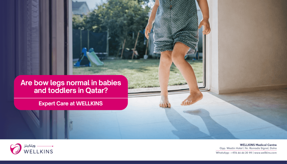
Are bow legs normal in babies and toddlers?
Yes, bow legs are entirely normal and expected in infants and toddlers. Most babies are born with a degree of bowing due to their position in the womb and this typically resolves naturally by age two to three as the child begins to walk and the legs straighten progressively. Bow legs that persist significantly beyond age three, that are worsening rather than improving, that are asymmetric with one leg more bowed than the other or that are accompanied by short stature warrant an orthopedic assessment to exclude an underlying condition.
When do knock knees in children correct themselves?
Knock knees, where the knees angle inward and the ankles are apart when the child stands straight, are very common in children between three and six years of age and typically correct naturally by around age seven to eight as the leg alignment continues its developmental progression. Knock knees that are still marked beyond age eight, that are causing pain or gait difficulty, that are significantly asymmetric or that are progressing rather than improving require specialist evaluation to determine whether an underlying cause and specific treatment are needed.
Can bow legs or knock knees cause long-term problems?
If left untreated when treatment is indicated, significant bow legs and knock knees can lead to early onset arthritis in the knee joint due to abnormal mechanical loading of the joint surfaces, chronic pain in the knee, hip or ankle, progressive deformity that becomes harder to correct as the child grows older and difficulty with walking and running. The key distinction is between mild self-resolving physiological alignment that causes no long-term issues and pathological alignment that does require management to prevent these complications.
What is guided growth surgery for bow legs and knock knees?
Guided growth surgery, known medically as hemiepiphysiodesis, is a minimally invasive surgical procedure that temporarily slows growth on one side of the growth plate to allow the natural growth process to gradually correct the alignment. It is most effective in children who still have significant growth remaining and produces excellent results with a relatively minor surgical procedure compared to corrective osteotomy. Small implants placed across one side of the growth plate are removed once the desired alignment has been achieved. It is one of the most elegantly simple interventions in paediatric orthopedic surgery.
1. What Are Bow Legs (Genu Varum)?
Bow legs occur when the knees curve outward, creating a visible gap between the lower legs when the child or adult stands with their feet together. From the front the legs appear to form a curve like the letter O rather than being vertically aligned. In infants and toddlers this pattern is entirely expected. In older children and adults it warrants clinical evaluation.
Causes of Bow Legs
- Physiological (Normal in Infants): Most babies are born with bow legs due to their position in the uterus during fetal development. This physiological bowing typically resolves naturally and progressively by age two to three without any intervention as the child begins walking and the mechanical loading of the limbs encourages natural alignment. No treatment is required or appropriate for physiological bow legs.
- Vitamin D Deficiency (Rickets): Inadequate vitamin D leads to poor calcium absorption and impaired bone mineralisation, producing soft bones that bow under the weight of the child’s body. In Qatar where sun avoidance due to intense heat and significant indoor time can contribute to vitamin D insufficiency particularly in certain communities, rickets-related bow legs are a clinically relevant consideration. Treatment addresses both the nutritional deficiency and the bony deformity.
- Blount’s Disease: A growth disorder specifically affecting the inner aspect of the upper tibia, the shin bone, that causes progressive worsening bowing rather than the natural resolution expected with physiological bowing. Blount’s disease is distinguished from physiological bowing by its persistence beyond age three, its tendency to be asymmetric and its progressive course. Early identification allows management with bracing in younger children or surgery in more established cases.
- Trauma or Infection: Damage to the growth plate from an injury or from infection during childhood can cause asymmetric or abnormal bone development that results in progressive bowing of the affected limb.
- Osteoarthritis: In adults, wear and tear arthritis of the medial knee compartment can progressively worsen an existing bowing tendency as cartilage loss on the inner side of the joint allows the leg to curve further outward.
- Genetic Conditions: Skeletal dysplasias including achondroplasia affect bone growth and development in ways that produce characteristic limb alignment patterns including bow legs. These conditions are identified through clinical and genetic assessment and require specialist management.
Symptoms of Bow Legs
- A visible outward curvature of the legs when standing with feet together.
- An awkward walking pattern, sometimes described as a waddling gait, particularly prominent in more significant deformities.
- Knee or hip pain in older children and adults where the abnormal alignment creates uneven mechanical loading of the joint surfaces.
- Uneven shoe wear, particularly on the outer aspect of the sole, reflecting the altered mechanical axis of the lower limb during walking.
2. What Are Knock Knees (Genu Valgum)?
Knock knees are the opposite alignment pattern to bow legs. The knees angle inward toward each other while the ankles remain apart when the child or adult stands straight, creating an appearance where the inner aspects of the knees touch or nearly touch. From the front the legs appear to form a curve like the letter X.
Causes of Knock Knees
- Physiological (Common in Children Ages Three to Six): Just as bow legs are normal in infants, knock knees are a normal and expected part of lower limb alignment development in children between approximately three and six years of age. The lower limb goes through a progressive alignment sequence from bowed at birth through neutral to mildly knock-kneed in early childhood before settling into adult alignment by approximately age seven to eight. This physiological knock knee pattern requires no treatment.
- Obesity: Excess body weight places increased stress on developing bones and growth plates and is a recognised contributing factor to the development and persistence of pathological knock knees. In Qatar where childhood overweight and obesity rates are significant, this is a particularly relevant consideration in the clinical assessment of a child with persistent knock knees.
- Injury or Infection: Growth plate damage from trauma or infection can cause asymmetric growth that produces a progressive knock knee deformity in the affected limb.
- Renal Disorders: Kidney disorders affect the metabolism of calcium and phosphate in ways that can impair bone mineralisation and produce knock knee deformity alongside other skeletal changes.
- Genetic Disorders: Conditions including Morquio syndrome and osteogenesis imperfecta affect skeletal development and can cause significant lower limb alignment problems that require specialist orthopedic management.
Symptoms of Knock Knees
- The knees touching or nearly touching while standing with the feet shoulder-width apart.
- Tripping or clumsiness, particularly common in younger children where the inward-angled knees can interfere with the normal walking pattern.
- Knee or ankle pain, particularly in older children and adults where abnormal mechanical loading of the outer knee compartment and ankle creates cumulative discomfort with activity.
- Difficulty running or walking smoothly due to the altered lower limb mechanics.
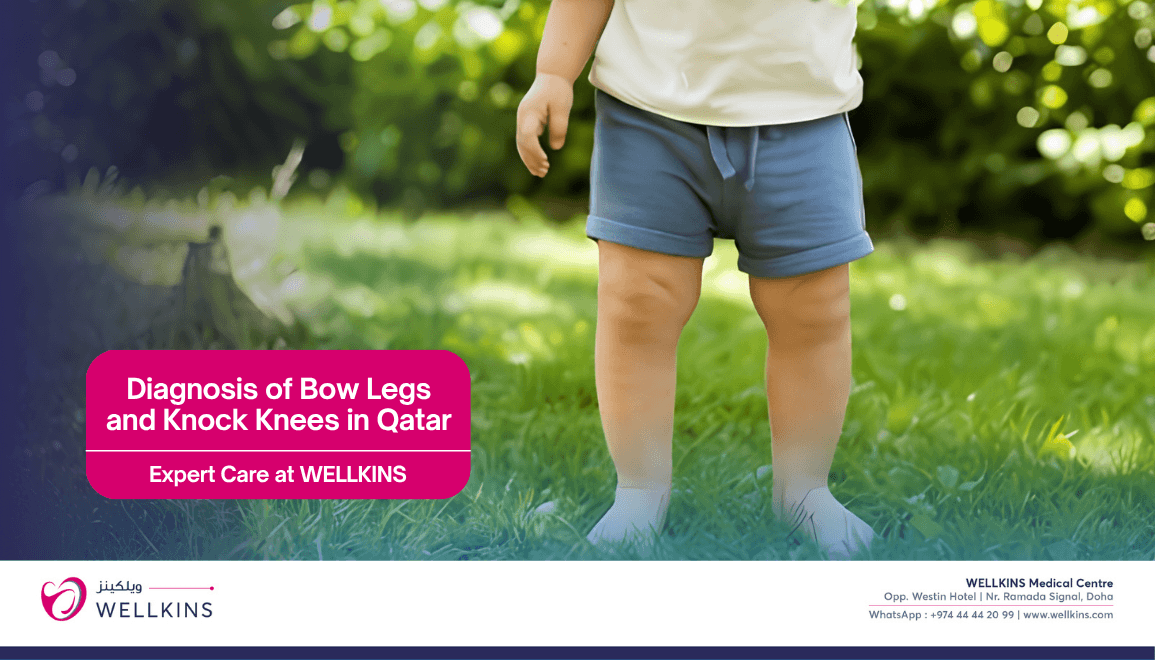
3. Diagnosis of Bow Legs and Knock Knees
At Wellkins Medical Centre the assessment of lower limb alignment uses a structured combination of clinical and imaging tools to determine whether the alignment is physiological or pathological and to identify any underlying cause.
- Physical Examination: Assessing the child’s or adult’s gait in the clinic corridor, measuring the intermalleolar distance for knock knees and the intercondylar distance for bow legs, examining the flexibility and range of motion of the hips, knees and ankles and evaluating the overall lower limb alignment in standing and walking positions.
- X-Rays: Long-leg standing X-rays that capture the entire lower limb from hip to ankle allow precise measurement of the mechanical axis deviation, the angle between the femur and the tibia and the specific location of the deformity within the limb. These measurements guide treatment decisions and provide a baseline for monitoring progression over time.
- Blood Tests: Ordered when a metabolic disorder including rickets or a renal condition is suspected as the underlying cause of the alignment problem. Vitamin D levels, calcium, phosphate and alkaline phosphatase are the most relevant markers in this context.
- MRI or CT Scans: Rarely required for straightforward alignment assessment but occasionally needed when growth plate abnormalities, Blount’s disease staging or preoperative planning for corrective surgery requires detailed imaging of the bone and growth plate structures.
4. Potential Complications If Untreated
When significant bow legs or knock knees are left without appropriate management the following complications can develop over time.
- Early Arthritis: Abnormal lower limb alignment concentrates the mechanical load of walking and standing onto specific areas of the knee joint surface. Over years this localised overloading accelerates cartilage wear and leads to early onset osteoarthritis that is considerably harder to manage than the original alignment problem would have been.
- Chronic Pain: Persistent knee, hip or ankle discomfort resulting from the abnormal mechanical loading patterns that accompany uncorrected alignment deformities, often becoming the primary driver of treatment requests in adults with longstanding untreated alignment problems from childhood.
- Mobility Issues: Difficulty walking or running efficiently due to the altered mechanics of the lower limb, limiting physical activity and quality of life.
- Progressive Deformity: Without intervention in cases where the underlying cause is not self-limiting, the deformity can worsen over time rather than plateauing, particularly during periods of rapid skeletal growth.
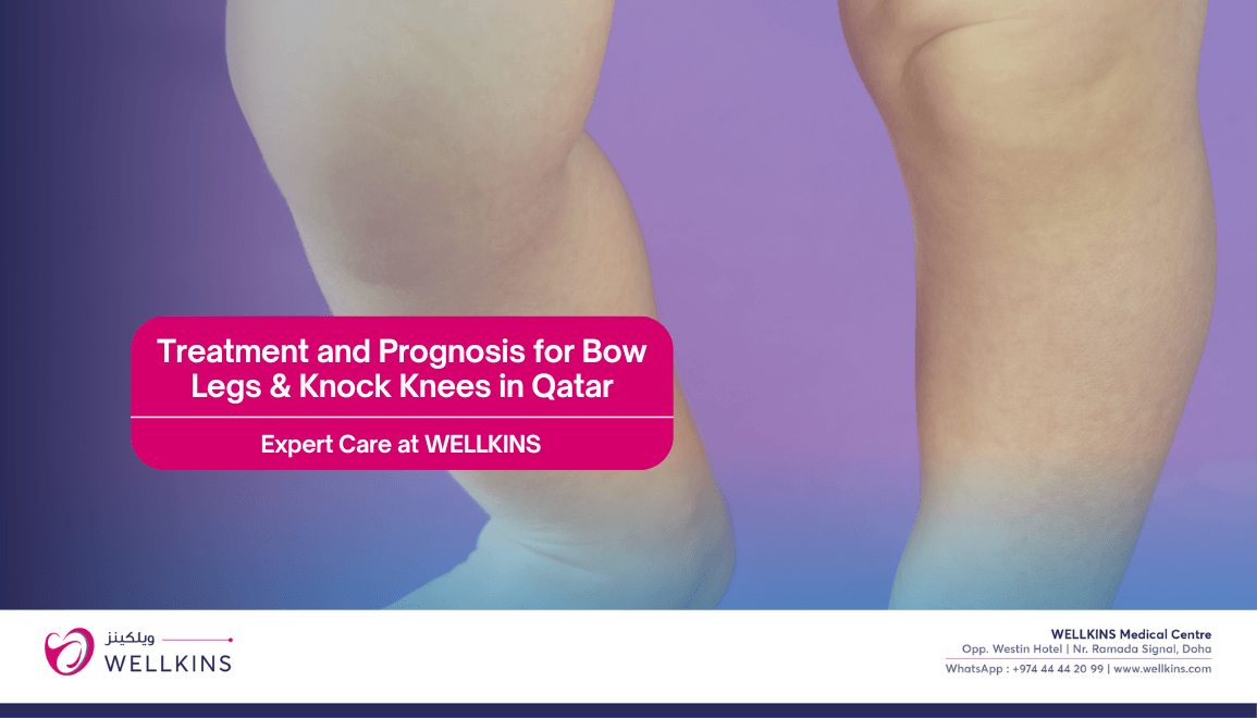
5. Treatment and Prognosis
Treatment decisions are guided by the severity of the deformity, the age of the patient, the underlying cause and whether the condition is progressing or stable.
Non-Surgical Management
- Observation: The most appropriate management for the majority of childhood cases. Many physiological alignment patterns resolve naturally through the normal developmental progression and require only periodic monitoring to confirm that resolution is occurring as expected.
- Vitamin D and Calcium Supplementation: For cases caused by rickets, correcting the nutritional deficiency is the primary treatment and often produces significant improvement in the bony alignment as the bone remineralises and strengthens during continued growth.
- Bracing: Used in early Blount’s disease in young children and in selected cases of moderate deformity to guide alignment during the growth phase. Bracing is most effective when initiated early and requires consistent use to produce benefit.
- Physical Therapy: Strengthening the muscles around the hip, knee and ankle improves the dynamic alignment of the lower limb during movement and reduces the joint pain associated with alignment disorders in older children and adults.
Surgical Options for Severe Cases
- Guided Growth Surgery (Hemiepiphysiodesis): A minimally invasive procedure that temporarily slows growth on one side of the growth plate to allow the natural growth process to progressively correct the alignment. This elegant approach harnesses the body’s own growth to produce correction and is most effective in children with significant growth remaining. Small implants are placed and subsequently removed once the desired alignment has been achieved.
- Osteotomy: A procedure in which the bone is surgically cut and realigned into the correct position and secured with plates and screws. Osteotomy is used in adolescents and adults in whom the growth plates are closed and guided growth is therefore no longer possible. It produces excellent and lasting correction but requires a longer recovery and rehabilitation period than guided growth surgery.
- Joint Replacement: In adults with severe arthritis resulting from longstanding untreated alignment deformity, joint replacement combined with alignment correction may be required to address both the mechanical and the degenerative components of the condition.
Prognosis
- Children: Most mild cases of physiological bow legs and knock knees resolve completely without treatment and leave no lasting impact on lower limb function or joint health.
- Adolescents and Adults: Surgical management has excellent outcomes when performed before severe joint damage has occurred. The earlier the intervention in pathological cases the better the long-term prognosis for both alignment and joint health.
Bow legs and knock knees are common conditions and for the majority of families the story ends reassuringly with natural resolution during childhood. For those cases that do require attention, early evaluation keeps the most effective and least invasive treatment options available and produces outcomes that protect the joints for a lifetime.
If your child or you has persistent leg alignment concerns beyond the expected developmental age, the right time to seek specialist assessment is now rather than later.
To book an appointment with Dr. Reneesh at Wellkins Medical Centre: https://wellkins.com/drreneesh
To know more about the Orthopedic services at Wellkins Medical Centre: https://wellkins.com/orthopedics